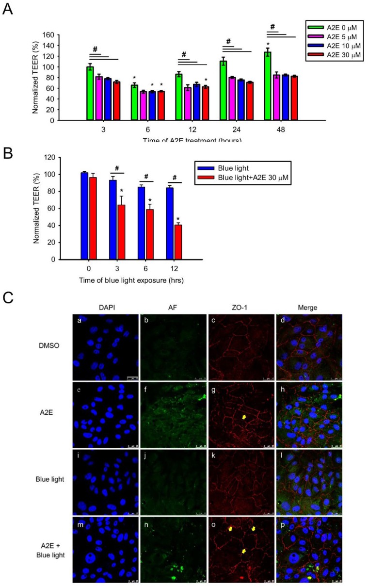
Figure 3.
A2E treatment increases paracellular permeability and blue light diminishes epithelial tight junction integrity in A2E-laden RPE cells. (A) The effects of osmotic stress on retinal epithelial barrier function are shown. The vertical bars show changes in resistance (Ω*cm2) of A2E-loaded RPE cells via trans-epithelial electrical resistance (TEER)measurements. RPE cells were treated with different concentrations of A2E (0, 5, 10, and 30 μM) for the indicated incubation time (3, 6, 12, 24, and 48 h). Results from 4 independent experiments are shown (N = 4). Data are presented as the normalized percent of average TEER ± SE. * p < 0.05 compared with the control group treated with same concentration of A2E; # p < 0.05 compared with the 0 μM A2E group during the same treatment time. (B) RPE cells were treated with 30 μM A2E and co-treated with different time of blue light-emitting diode (LED) light (BLL) exposure (460 nm, 150 lux; 0, 3, 6 and 12 h). Data are presented as the normalized percent of average TEER ± SE. * p < 0.05 compared with the control group treated with 0 h BLL exposure; # p < 0.05 compared with the BLL group. (C) Photographs showing A2E autofluorescence (green) and ZO-1 immunofluorescence staining (red) are depicted. Cell nuclei were stained with 4′-6-diamidino-2-phenylindole (DAPI, blue). RPE cells were treated with 30 μM A2E and subjected to BLL for 12 h (460 nm, 150 lux). BLL exposure resulted in the formation of discontinuous ZO-1-positive strands in A2E-laden RPE cells (as indicated by yellow arrows). Results from 3 independent experiments are shown (N = 3). Scale bar, 25 μm.

