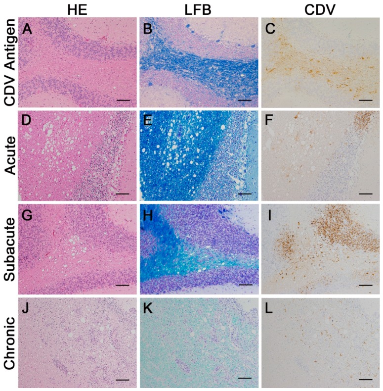
Figure 2.
Classification of cerebellar lesions in the white matter of canine distemper virus (CDV)-infected dogs. (A–C) Group 2: antigen expression without histological lesions. (D–F) Group 3: acute lesion. (G–I) Group 4: subacute lesion. (J–L) Group 5: chronic lesion with prominent inflammation. (A,D,G,J) hematoxylin and eosin (HE)-staining; (B,E,H,K) luxol fast blue (LFB) staining; (C,F,I,L) CDV. Immunohistochemistry visualized by the avidin-biotin-peroxidase complex (ABC) method with 3,3′-diaminobenzidine as substrate and counterstaining with Mayer’s hematoxylin. Bars, 100 µm.

