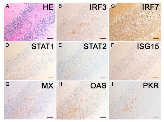
Figure 4.
IRF3, IRF7, STAT1, STAT2, ISG15, MX, OAS and PKR protein expression in serial sections of a chronic white matter lesion in the cerebellum of a canine distemper virus-infected female mixed breed dog. (A) Lesion with perivascular inflammatory cells (hematoxylin and eosin (HE)-staining); (B) Strong IRF3 protein expression of glial cells and perivascular lymphocytes; (C) Strong IRF7 staining of neurons and glial cells; (D) STAT1 protein expression of glial cells and a few neurons; (E) Strong immunostaining of glial cells and Purkinje cells for STAT2 proteins; (F) ISG15 protein expression of granular cell layer neurons and intralesional glial cells; (G) Strong Mx protein expression of numerous intralesional glial and inflammatory cells as well as Purkinje cells; (H) Strong OAS protein expression of perivascular lymphocytes, intralesional glial cells and Purkinje cells; (I) Strong PKR protein expression particularly of intralesional lymphocytes. Immunohistochemistry visualized by the avidin-biotin-peroxidase complex method with 3,3′-diaminobenzidine as substrate and counterstaining with Mayer’s hematoxylin. Bars, 100 µm.

