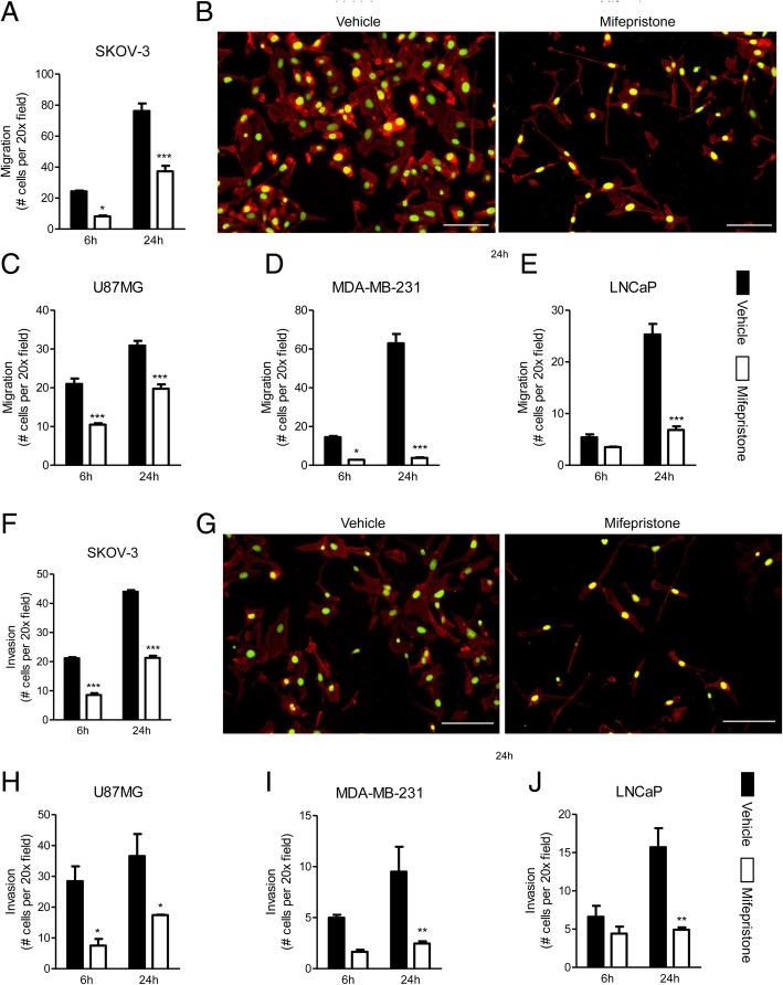
Fig. 4.
Mifepristone reduces the migration and invasion of cancer cells of the ovary (a, b, f, g), glia (c, h), breast (d, i), or prostate (e, j) in a Boyden chamber assay assessed by fluorescence microscopy. Each cell line was treated with their respective concentration of MF for 72 h prior to plating. Visual representations of migrated (b) and invasive (g) SKOV-3 cells, labeled with Alexa Fluor® 594-phalloidin and SYTOX® Green Nucleic Acid stain upon treatment with vehicle (left panels) or MF (right panels). Scale bar = 100 μm. Data shown represent the mean ± s.e.m. Vehicle (closed bars); MF (open bars). *P < 0.05, **P < 0.01, ***P < 0.001 compared against vehicle. Statistical analysis was done using two-way ANOVA followed by Bonferroni’s multiple-comparison test

