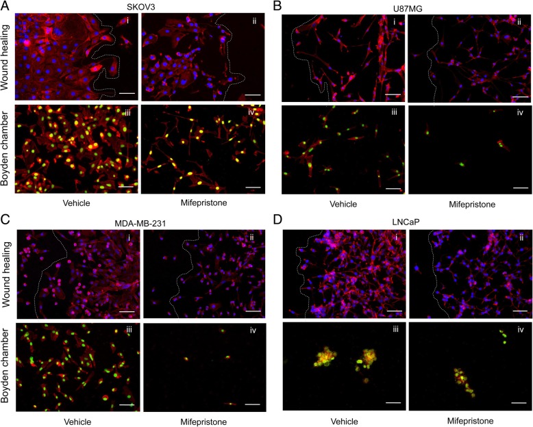
Fig. 5.
Enhancing the wound healing and Boyden chamber assays with a double fluorescence labeling allows for the visualization of different migration patterns between cell lines. SKOV-3 (a), U87MG (b), MDA-MB-231 (c), or LNCaP (d) were all treated with their respective concentrations of MF for 72 h. A wound healing assay and a Boyden chamber assay were performed for each cell line. After 24 h, cells were fixed with 4% PFA, and stained with Alexa Fluor®-594 Phalloidin and DAPI (wound healing), or SYTOX® Green Nucleic Acid stain (Boyden chamber). Scale bars = 75 μm. White lines represent the front of the wound

