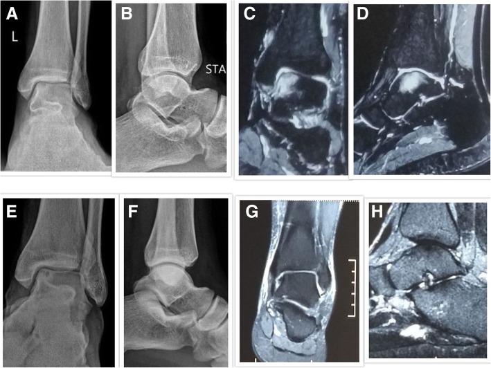
Fig. 1.
a, b Pre-treatment anteroposterior and lateral radiographs of a 50-year-old male diagnosed with modified Ficat and Arlet stage II avascular necrosis of the talus bone. c, d Pre-treatment MRI Coronal and Sagittal images showing BME. e, f Post-treatment anteroposterior and lateral radiographs at 50 months follow-up showing no radiological collapse/progression with stabilization of the AVN in stage II. g, h Post-treatment MRI coronal and sagittal images showing complete resolution of BME

