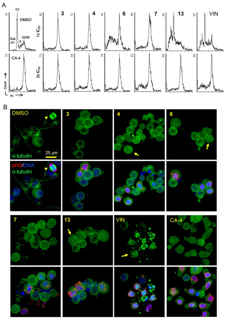
Figure 2.
Mitotic defects in KB-VIN cells treated by compounds. (A) Vincristine-resistant subline KB-VIN cells were treated with compounds for 24 h at a concentration of one- or three-fold IC50 (1× IC50 or 3 × IC50). CA-4 at 0.2 µM was used as a colchicine-type tubulin polymerization inhibitor. Cell cycle distributions (sub-G1, G1, S, G2/M) were analyzed using flow cytometry after staining cells with propidium iodide (PI). (B) KB-VIN cells were treated with compounds for 24 h at a concentration of 3 × IC50. CA-4 was used at 0.2 μM. Fixed cells were stained with antibodies to α-tubulin (green) and phospho-histone H3 (pH3, red), and DAPI was used for DNA (blue). Stained cells were observed by confocal fluorescence microscope. The represented image is a projection of 15~20 optical sections acquired at 0.5~1 µm intervals. Normal mitotic spindle formation (arrow head) in control (DMSO) and dotted tubulin aggregations without spindles (4, 6, 13) or with multipolar spindles (VIN) were observed (arrows). Bar, 0.025 mm. Additional images are available in Supplementary Figure S1.

