Fig. 3.
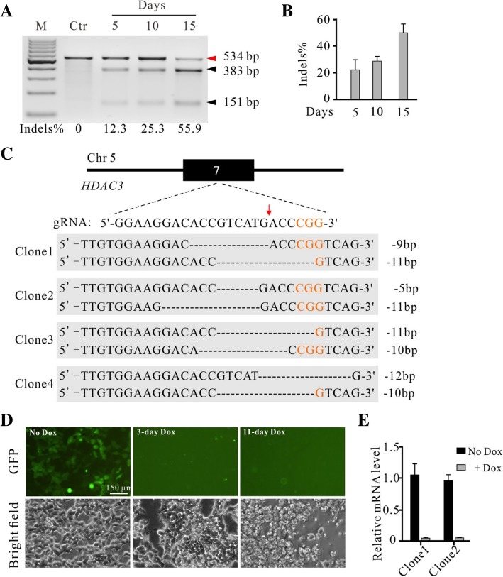
Generation of HDAC3 knockout-rescue cell lines. a Representative gel pictures of T7E1 assay for detection of indels at HDAC3 sites in HEK293T cells. Ctr is the PCR band from unmodified cells with T7 enzyme digestion. b Quantification of the T7E1 assay for Fig. 3a (n = 3, error bars show mean ± SEM). c Examples of indel sequences for four single cell-derived clones. Schematic of the gRNA target site was shown above. PAM sequence was marked in orange. Cas9 cutting site is indicated by red arrow. d Inhibition of exogenous HDAC3 expression in HDAC3-knockout cells caused cell death. The HDAC3 knockout-rescue cells expressed GFP. Expression of GFP was inhibited by addition of Dox for 3 days. All cells died at day 11. e RT-qPCR analysis of the exogenous HDAC3 expression with or without Dox for two clones (n = 3, error bars showed mean ± SEM)

