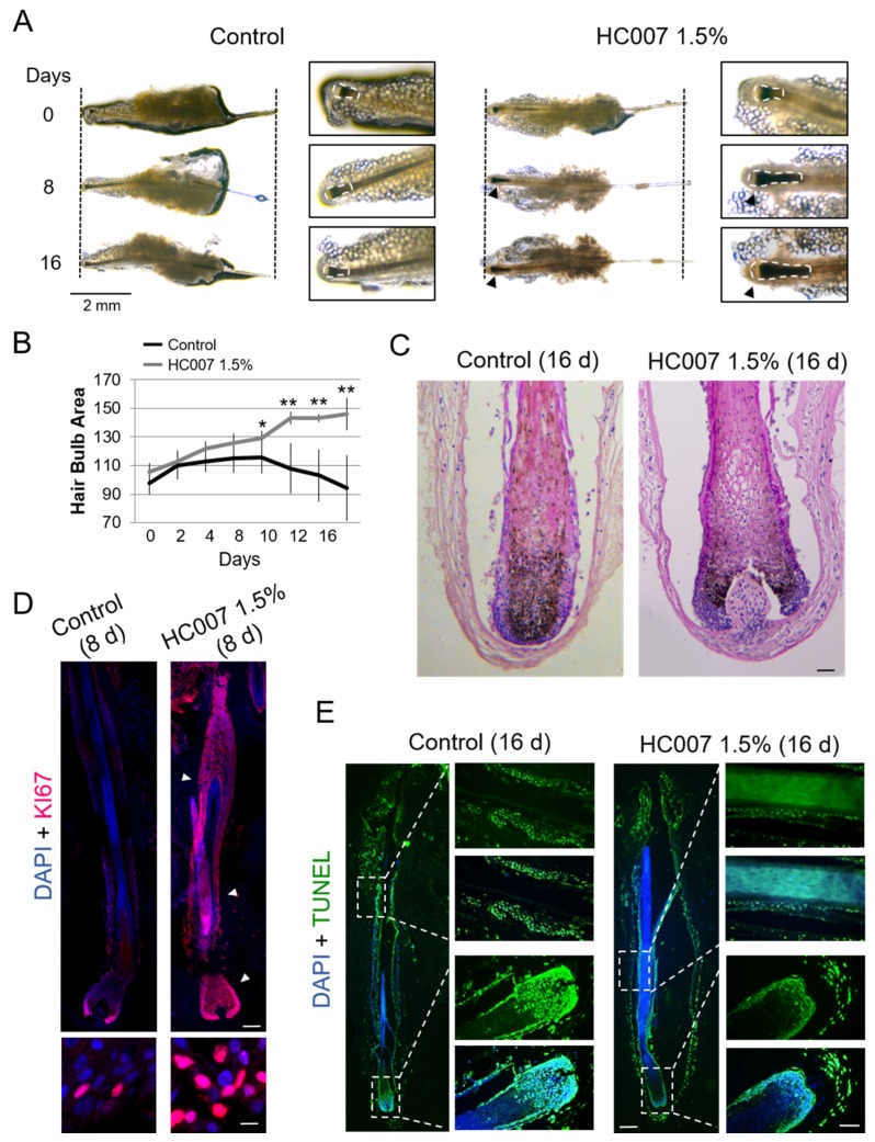
Figure 1.
Sustained maintenance of human hair follicle growth ex vivo in a defined glycosaminoglycan hydrogel matrix (HC007). (A) Representative phase-contrast microscopy images of whole hair follicles showing significant thickening (black arrowheads) of the hair bulb area, defined here as an area encompassing hair bulb/dermal papilla and suprabulbar regions, after continuous growing in HC007 1.5%. Vertical dotted lines delimitate hair follicle length at time 0. A noticeable length increase in hair follicle length was observed in 50% of the HC007 1.5% samples. Enlarged images depicting typical hair bulb areas used in image quantification analysis (white dotted lines). Bar: 2 mm. (B) Time course quantification of hair bulb area in control and HC007 1.5% samples. Results are representative of at least 18 hair follicle units per condition. The mean +/− SD of n ≥ 18 for each experimental condition is represented and t-test was used for statistical analysis. *, significant p ≤ 0.1. **, significant p ≤ 0.05. (C) Representative histological sections stained with H&E of the hair bulb area in control and HC007 1.5% after 16 days in culture. Bar: 50 µm. (D) Confocal microscopy images (maximum projections) showing the localization of the cell proliferation marker KI67 in morphologically equivalent histological sections of hair follicles grown ex vivo for 8 days in control basal culture conditions or in HC007 1.5%. Bar: 100 µm. Enlarged images show nuclear KI67 staining in outer root sheath cells. Bar: 10 µm. (E) Identification of apoptotic cells by the TUNEL assay in morphologically equivalent histological sections of hair follicles grown ex vivo for 16 days in control basal culture conditions or in HC007 1.5%. Bars: 100 µm. Results shown in (C–E) are representative of at least 10 hair follicles in three independent experiments.

