Figure 1.
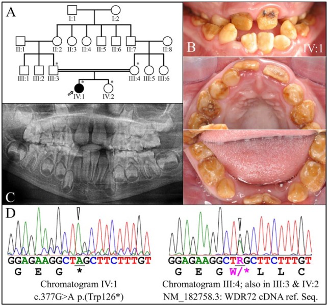
Family 1. (A) Pedigree of family 1. The proband (IV:1) is the only affected member and is indicated by an arrow. The asterisks (*) indicate the 4 family members recruited for this study. The double line connecting the father (III:3) and the mother (III:4) of the proband indicates consanguinity. (B) Clinical photos of the proband’s dentition in the mixed dentition stage (age, 8 y). Hypomaturation amelogenesis imperfecta is evident in the primary and secondary dentitions, which were stained brown and had undergone rapid attrition. Black stains on the maxillary permanent central incisors suggested the early onset of dental caries. (C) Panoramic radiograph of the proband. The enamel layer was barely visible radiographically, although the sizes and shapes of the crowns were within normal limits at the time of eruption, suggesting that enamel layer thickness was within normal limits. (D) DNA sequence chromatograms confirmed that the proband was homozygous for the WDR72 c.377G>A mutation that introduced a p.(Trp126*) stop gain in exon 5 (left). The mother (III:3), father (III:4), and sister (IV:2) of the proband were all heterozygous for this defect (right).

