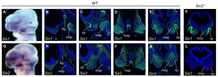
Figure 1.
Expression of Six1 and Six2 in the developing frontonasal, maxillary, and mandibular processes. (A) Whole-mount view of the pattern of Six1 messenger RNA (mRNA) expression in E10.5 wild-type (WT) embryo. (B–E) Representative sagittal (B, C) and frontal (D, E) sections of E10.5 WT embryos showing immunofluorescent staining (green) using the anti-SIX1 antibody. DAPI counterstaining is shown in blue. (F) Representative frontal section of E10.5 Six2–/– embryo showing immunofluorescent staining (green) using the anti-SIX1 antibody. (G) Whole-mount view of the pattern of Six2 mRNA expression in E10.5 WT embryo. (H–K) Representative sagittal (H, I) and frontal (J, K) sections of E10.5 WT embryos showing immunofluorescent staining (green) using the anti-SIX2 antibody. (L) Immunofluorescent staining of frontal sections of E10.5 Six2–/– embryos using the same anti-SIX2 antibody. Multiple serial sections from 3 wild-type and 3 Six2–/– embryos were used for immunofluorescent staining using each antibody. hm, head mesenchyme; mdp, mandibular process; mnp, medial nasal process; mxp, maxillary process; np, nasal pit; Rp, Rathke’s pouch. Scale bar in panel B, 100 µm.

