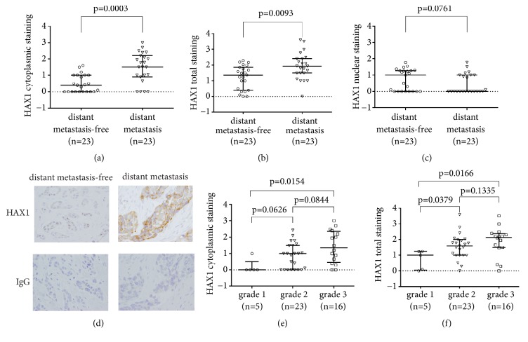
Figure 4.
HAX1 protein level in primary tumors stratified according to selected clinical and histological factors (presence of distant metastases, tumor grade). (a-d) HAX1 protein levels in the primary tumor were quantified from IHC data in distant metastasis-free versus distant metastasis group for (a) cytoplasmic, (b) total, and (c) nuclear HAX1 staining. (d) Representative images of HAX1 IHC and negative isotype control for patients from metastasis-free versus distant metastasis group. ×40 objective, bar: 100 μm. (e) Cytoplasmic and (f) total HAX1 staining in breast cancers stratified according to tumor grade (grades 1-3). Results for individual patients and median and interquartile range for each group are shown. Differences in HAX1 protein levels between groups were assessed by the Mann-Whitney U test and results with p-values <0.05 were considered significant.

