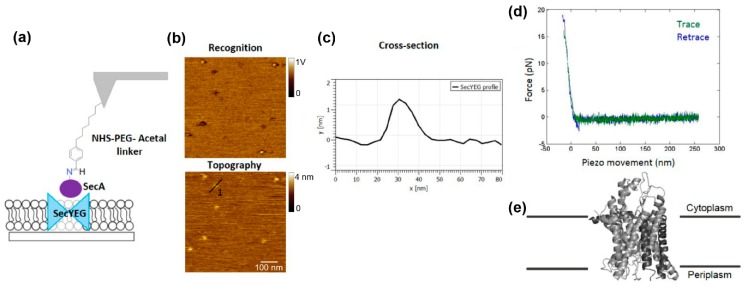
Figure 4.
Proof of concept: characterization of the SecA–SecYEG interaction. (a) Scheme of SecYEG complexes reconstituted into a planar lipid bilayer with the tip carrying a SecA molecule attached via an N-hydroxysuccinimide (NHS)–PEG–Acetal linker. (b) SecYEG molecules that interacted with the SecA on the tip appear as dark spots in the recognition images. (c) Cross-section of SecYEG. (d) Characteristic force-distance curve for the SecYEG–SecA interaction. (e) Side view of the crystal structure of SecYEG from M. Jannaschii (PDB-ID 1RHZ).

