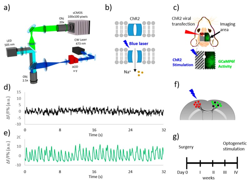
Figure 1.
Double-split illumination path. (a) Schematic representation of wide-field microscope for the simultaneous laser stimulation and cortical imaging. (b) Schematic representation of Channel rhodopsin 2 structure. (c) Schematic representation of field of view for inter-hemispheric optogenetic investigation in GCaMP6f mice: ChR2-transfected M1 (red circle) in the left obscured hemisphere is stimulated by the 473 nm blue laser; cortical activity is revealed in the right hemisphere. (d) Left hemisphere ΔF/F signal during resting state; (e) Right hemisphere ΔF/F signal during resting state; (f) Schematic cartoon of interhemispheric connectivity of homotopic regions of the brain cortex via corpus callosum. The red spots represent ChR2-transfected neurons, the green spots represent excitatory neurons expressing the calcium indicator GCaMP6f, the black spots represent inhibitory neurons. (g) Experimental timeline. AOD: Acousto-optic deflector.

