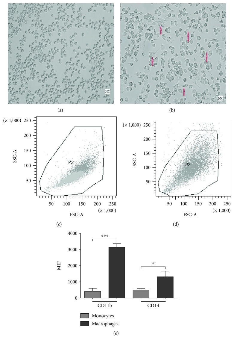
Figure 1.
Induction of the macrophage-like phenotype in THP1 cells. THP-1 cells were differentiated by PMA treatment. Morphological changes were analyzed by comparing (a) undifferentiated cells versus (b) differentiated cells. Arrows indicate typical “spreading.” Differentiation was corroborated by analyzing the increment in size and granularity using flow cytometry, comparing (c) nondifferentiated vs. (d) differentiated cells. (e) PMA-treated cells showed increased levels of CD11b and CD14 expression. ∗∗∗ P ≤ 0.0001.

