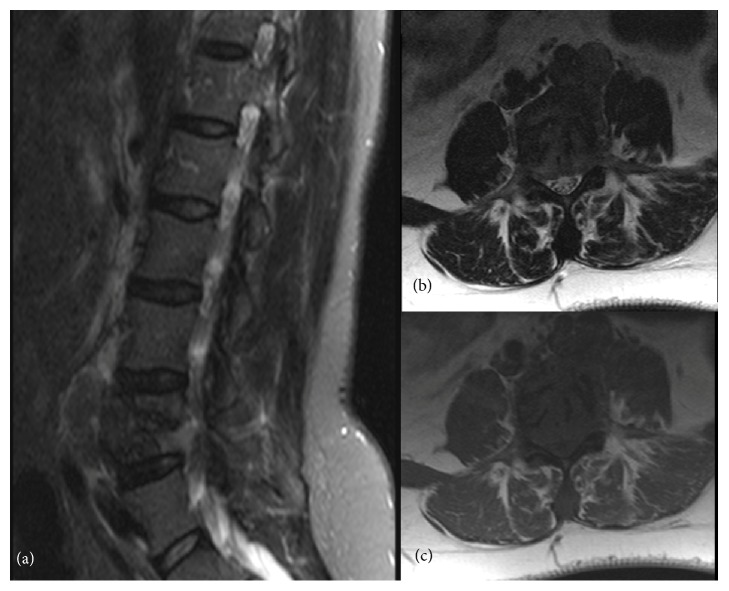
Figure 1.
MR of the lumbar spine. (a) T2-weighted sagittal Spectral Attenuated Inversion Recovery (SPAIR) image. (b) T1-weighted axial image. (c) T2-weighted axial image. There is a T1-hypointense, T2-heterogeneous soft tissue mass occupying and destroying a large part of L4, causing nerve root compression.

