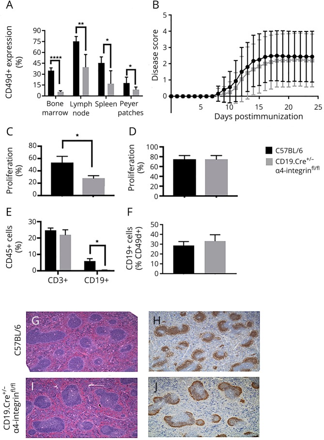
Figure 1. CD19.Cre+/− α4-integrinfl/fl mice are fully susceptible to actively MOGp35-55-induced experimental autoimmune encephalomyelitis.
(A) The percentage of CD49d+ CD19+ B cells in the bone marrow, lymph nodes, spleens, and Peyer patches is significantly diminished in naive CD19.Cre+/− α4-integrinfl/fl mice compared with C57BL/6 control mice. Cells were immunophenotyped by multiparameter flow cytometry. (B) CD19.Cre+/− α4-integrinfl/fl mice behave similar to C57BL/6 wild-type (WT) mice regarding the disease incidence, onset, and severity of active experimental autoimmune encephalomyelitis (EAE). α4-integrinfl/fl mice were used as controls and showed similar disease activity (data not shown). Active EAE was induced in CD19.Cre+/− α4-integrinfl/fl and C57BL/6 age-matched control mice by subcutaneous immunizations with MOGp35-55 in incomplete Freund adjuvant (IFA) containing 4 mg/mL mycobacteria. Mice received intraperitoneal injection of 200 μL of pertussis toxin (Ptx) at 200 ng/mL on days 0 and 2. Mice were observed daily, and EAE severity was scored using a 5-point scale. CD4+ T-cell recall responses to MOGp35-55 in (C) spleens were significantly diminished in CD19.Cre+/− α4-integrinfl/fl mice, but (D) indistinguishable to those in C57BL/6 WT mice in lymph nodes. Cell proliferation was determined by a flow cytometric proliferation assay using the green fluorescent dye CFSE or V450. (E) At maximum disease activity (days 13–15), the percentage of CD3+ T cells in the CNS was similar in CD19.Cre+/− α4-integrinfl/fl and C57BL/6 WT mice. In contrast, the percentage of CD19+ B cells in the CNS was significantly diminished in CD19.Cre+/− α4-integrinfl/fl mice. (F) The percentage of α4-integrin–positive (CD49d+) CD19+ B cells in the CNS during maximum EAE disease activity was comparable between both mouse strains. Lymphocytes were immunophenotyped by multiparameter flow cytometry. The number, appearance, and architecture of germinal centers in lymph nodes of (G and H) C57BL/6 WT control mice were comparable to those in (I and J) CD19.Cre+/− α4-integrinfl/fl mice. Panels G and I were stained with hematoxylin & eosin, and panels H and J were stained with anti-CD19. Magnification for G–J is ×4. *p < 0.05, **p < 0.01, ***p < 0.001, ****p < 0.0001. CFSE = carboxyfluorescein succinimidyl ester.

