Figure 2.
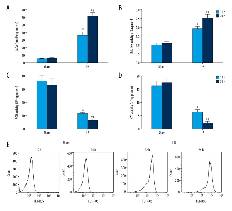
The oxidative stress and apoptosis of myocardium in I-R rats increased significantly. (A) Detection of MDA content in rat myocardium. (B) Caspase-3 activity in rat myocardium was detected by spectrophotometry. (C) SOD activity in rat myocardium was detected by kits. (D) CAT activity in rat myocardium was detected by kits. (E) Flow cytometry was used to detect ROS content in rat myocardium. * P<0.05 compared with the sham group; # P<0.05 at 24 h compared with 12 h.

