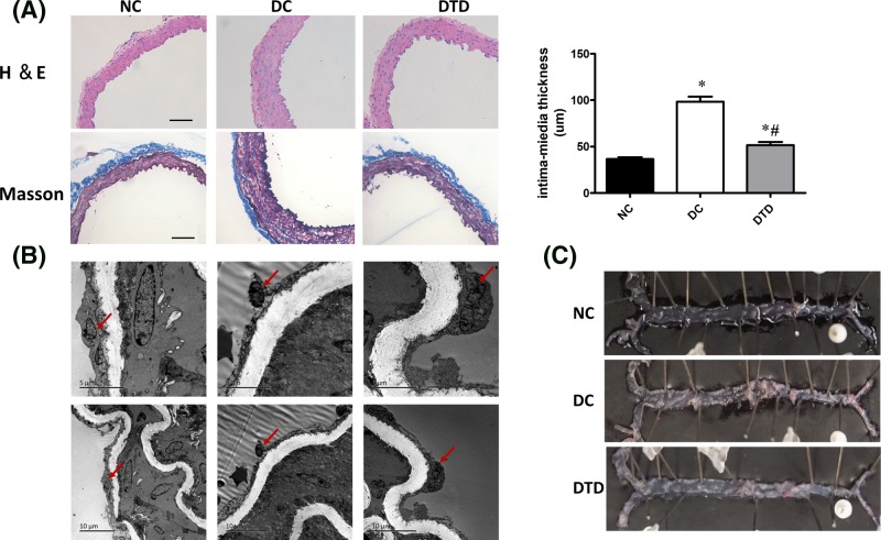
Figure 3. Desipramine treatment improved the morphological changes in aortas of db/db mice.
(A) Aortic histological sections were stained with H&E and trichrome. Desipramine treatment decreased the intima-media thickness of aortas from db/db mice. Original magnification is 200×. Scale bars represent 100 μm. (B) The endothelium structure changed as determined by electron microscopy observation among the three groups. Endothelial cells (bold arrow). Desipramine treatment prevented endothelial denudation of aortas from db/db mice. (C) Examination of the lipid deposition stained with Oil Red O. Data are shown as the mean ± S.E.M. with n=5 animals per group. *P<0.05 compared with the NC group. #P<0.05 compared with the DC group.

