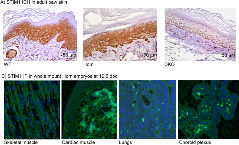
Figure 2. Stim1 expression in tissues from Stim1R304W mice.
(A) STIM1 IHC in adult paw skin demonstrates strong cytoplasmic STIM1 staining (brown) in epidermal keratinocytes in Stim1+/+ (WT, left), and Stim1R304W/R304W mice (Hom, middle). No staining was detected in the epithelial cell specific Stim1/2 double KO mouse (DKO, right) [39].
(B) STIM1 immunofluorescence (IF) in whole mount homozygous Stim1R304W embryos at 16.5 dpc demonstrates STIM1 staining (green) in cells of mesodermal origin (skeletal muscle cells and cardiac muscle cells), in cells of endodermal origin (lung cells), and in cells of ectodermal origin (cells in choroid plexus), as indicated.

