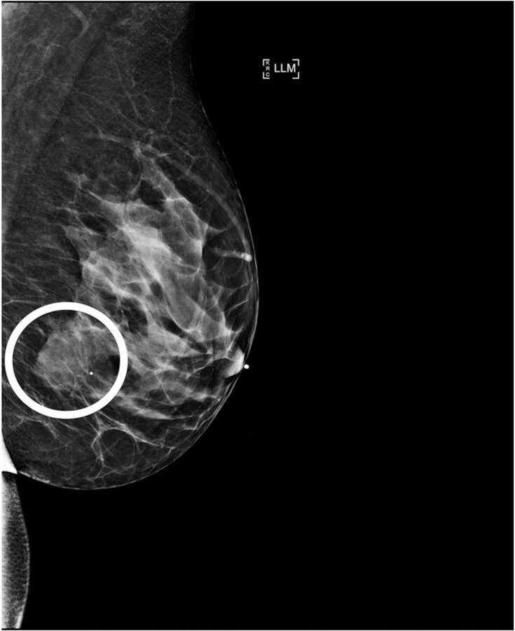
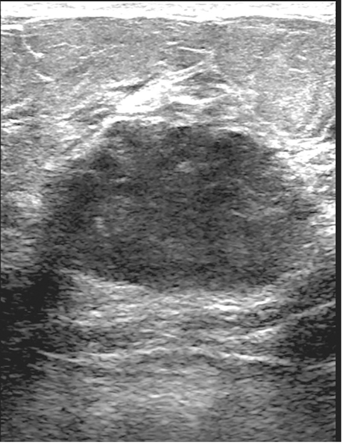
Figure 2.


38-year-old woman with AR– TNBC. (a) Left lateromedial mammogram shows an oval mass (white circle) without calcifications and with circumscribed margins. (b) Breast ultrasound of the same mass shows oval shape, circumscribed margins, and hypoechoic echo pattern. (c) Axial subtraction MR image of the same mass shows oval shape, circumscribed margins, and heterogeneous internal enhancement.
