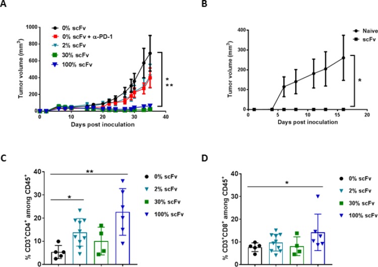
Figure 6. Pre-transduced tumor cells expressing scFv PD-L1 demonstrate a dose-dependent anti-tumor activity and enhanced tumor infiltrating lymphocytes.
(A) Subcutaneous tumor model using Tu-2449SC cells maximally infected with RRV-scFv PDL1 or RRV-GFP at indicated ratios were subcutaneously implanted on the right flank of 8-week-old female B6C3F1 mice and assigned to indicated groups (n = 10 per group). Control groups are mice implanted with 100% Tu-2449SC/RRVGFP cells that received PBS (0% scFv) or anti-mouse PD-1 antibody (0% scFv + α-PD-1). Anti-PD-1 antibody (Clone J43) was i. p. administered on day 0 (300 µg per mouse), day 3, day 6 and day 9 (200 µg per mouse). Statistical significance was determined by 2-way ANOVA of the following data sets: *p < 0.0001 for 0% vs 30% scFv; **p < 0.0001 for 0% vs 100% scFv. (B) Naïve mice (n = 5) and mice that cleared their initial tumor implant from RRV-scFv-PDL1 treated group (n = 15) were challenged with 2 × 10⁶ Tu-2449SC on the left side of the flank. Tumor growth and measurement were monitored over time. *p < 0.0001 (0% scFv vs. 2%, 30% and 100% scFv). Error bars indicate the standard error measurement (SEM) of the dataset. (C) and (D) Frequency of tumor-infiltrating CD4+ (C) and CD8+ (D) lymphocytes among CD45+ cells by flow cytometry, 39 days post-tumor implantation. Statistical analyses were performed using Mann-Whitney test at 95% CI (*P < 0.05, **P < 0.01). Values represent means ± SD of the dataset.

