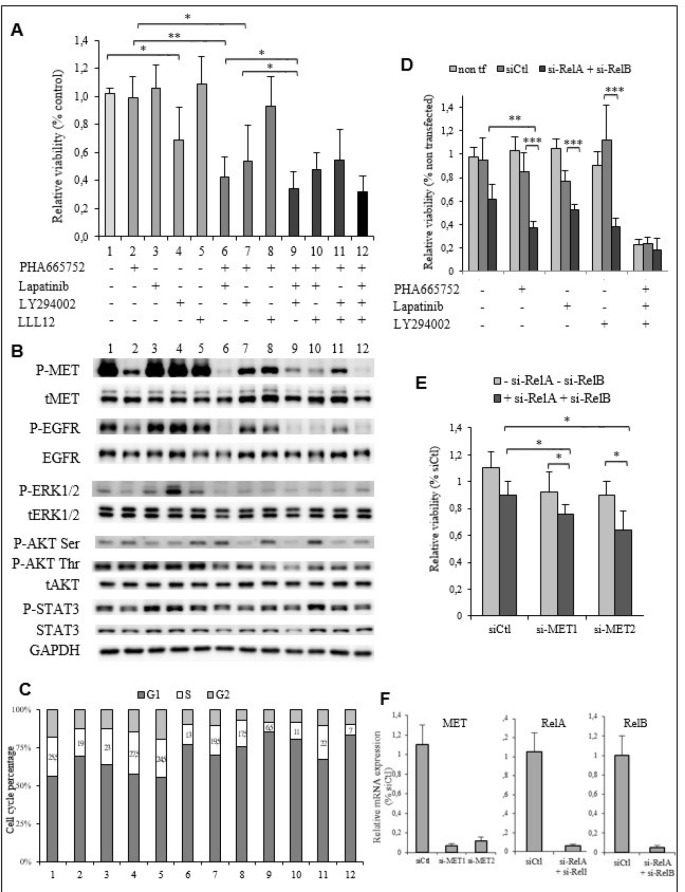
Figure 5. The EGFR, PI3K/AKT and NF-κB signaling pathways are implicated in PHA665752 resistance of the TTA1 cells.
(A) The TTA1 cells were incubated with the PHA665752, lapatinib, LY294002 and LLL12 inhibitors alone or in combination, as indicated. Cell viability was quantified by the crystal violet staining assay on day 3. (B) Western blot analysis of phosphorylated and total MET, EGFR, ERK1/2, AKT and STAT3 in TTA1 cells incubated with the combination of inhibitors used in viability assay reported in Figure 5A. (C) Cell cycle analysis after propidium staining of TTA1 cells incubated for 2 days with the various inhibitors combinations, as indicated in Figure 5A. (D) TTA1 cells were transfected with RelA si-RNA and RelB si-RNA and treated or not with PHA665752, lapatinib, and/or LY294002, as indicated, for 2 days. Cell viability was determined by using the crystal violet staining assay. The mean results of 8 replicates in two experiments are reported. (E) The TTA1 cells were transfected with the MET si-RNA1 and MET si-RNA2 with RelA si-RNA and RelB si-RNA or not. Cell viability was determined by using the crystal violet staining assay on day 3. The mean results of 8 replicates in two experiments are reported. (F) MET, RelA and ReB mRNA expression analysis by qRT-PCR in TTA1 cells 2 days after transfection with used in cell viability assay reported in Figures 5D–5E, as indicated.

