Figure 3.
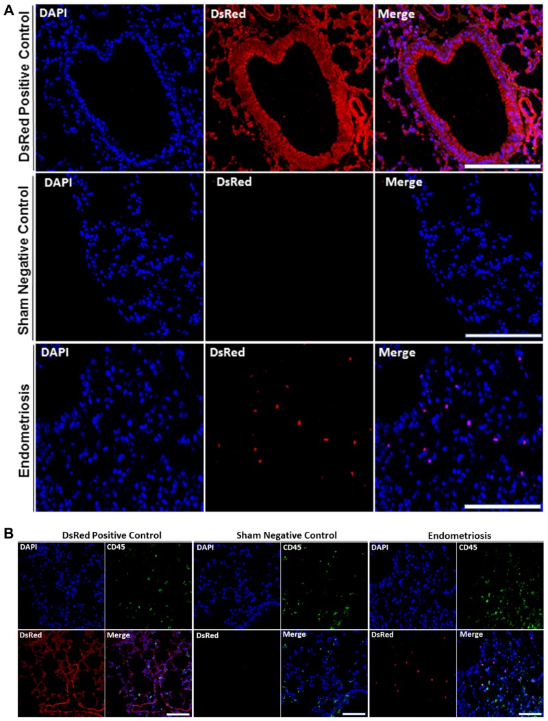
(A) DsRed+ cells are present in lung parenchyma of mice with endometriosis. Analysis of DsRed+ cell localization by confocal microscopy showing intense DsRed immunofluorescence in transgenic DsRed mice. No DsRed expression was observed in sham control mice. In each image, blue represents cell nuclei [4′, 6′- diamino-2-phenylindole (DAPI)], red indicates DsRed. Scale bars, 100 μm (Transgenic DsRed mouse and Sham), 50 μm (Endometriosis). Immunofluorescent images are representative of 3 random fields in each slide, with n = 10 mice in each group, in two independent experiments. (B) DsRed+ cells did not express CD45 in lung parenchyma of mice following induction of endometriosis by using double immunofluorescence. Confocal microscopic view of lung parenchyma. DsRed+/Cd45− cells were observed in the lung tissue of mice with experimental endometriosis. CD45 was used as a leukocyte marker to exclude migrating white blood cells. CD45+ cells (leukocytes) were observed, as expected, in all models including controls. Blue staining demonstrates cell nuclei [4′, 6′- diamino-2-phenylindole (DAPI)], while red staining corresponds to DsRed, and green staining shows CD45. Scale bars, 100 μm (Transgenic DsRed mouse and Sham), 50 μm (Endometriosis). The negative control tissues have no DsRed cells but have CD45+ leukocytes. The positive control DsRed tissues have all DsRed+ cells, including the CD45+ double positive leukocytes. The experimental mice have DsRed+ cells from the endometriosis that are not leukocytes and are therefore CD45−. Images are representative of three random fields in each slide, with n = 10 mice in each group, in two independent experiments.

