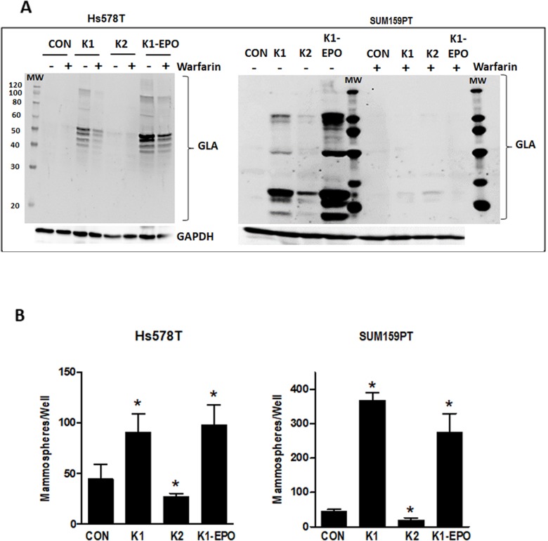
Figure 5. Effects of vitamins K2, K1-EPO, and VKOR inhibitor on GLA protein expression and mammosphere formation in TNBC cells.
(A) Hs578T (left) and SUM159PT (right) cells were maintained in media containing ethanol vehicle or 5 μg/ml K1, K2, or K1-EPO for > 3 passages. Post-attachment, cells were switched to media ± K1, K2, K1-EPO, and 2 μM warfarin for 48 h. Whole cell lysates were analyzed by western blotting for GLA or GAPDH as loading control. Blots are representative of at least 3 independent samples with similar results. (B) Hs578T (left) and SUM159PT (right) cells were grown > 3 passages in standard media containing EtOH vehicle or 5 μg/ml K1, K2, or K1-EPO and plated in ultra-low attachment plates in Mammocult™ media (STEMCELL Technologies). After 8 days, mammospheres were imaged and counted. Bars represent mean ± SD of 3 biological replicates analyzed in triplicate. *Significantly different from CON (p < 0.05) as measured by one-way ANOVA and Tukey post-test.

