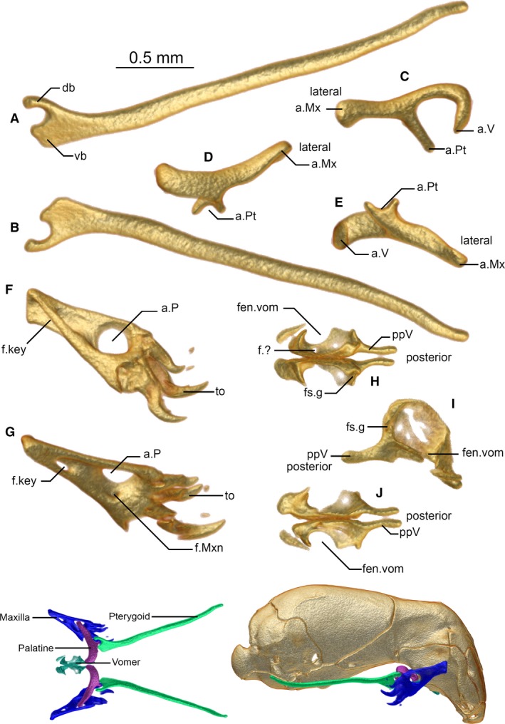
Figure 5.

Pterygoid (A,B), palatine (C–E), maxilla (F,G), and vomers (H–J) of Xenotyphlops grandidieri in (A,D,G,H) dorsal, (B,E,J) ventral, (C) anterior, (F) medial, and (I) lateral view. Colour insets show the articulated jaw complex in dorsal view (left) and articulated in lateral view (right). a.Mx, articulation of the palatine with the maxilla; a.P, articulation of the maxilla with the palatine; a.Pt, articulation of the palatine with the pterygoid; a.V, articulation of the palatine with the vomer; db, dorsal branch of the pterygoid; f.?, unknown foramen; f.key, keyhole foramen; f.Mxn, maxilla nerve foramen; fen.vom, fenestra vomeronasalis; fs.g, forsaken groove; ppV, posterior process of the vomer; to, tooth; vb, ventral branch of the pterygoid.
