
Figure 8.
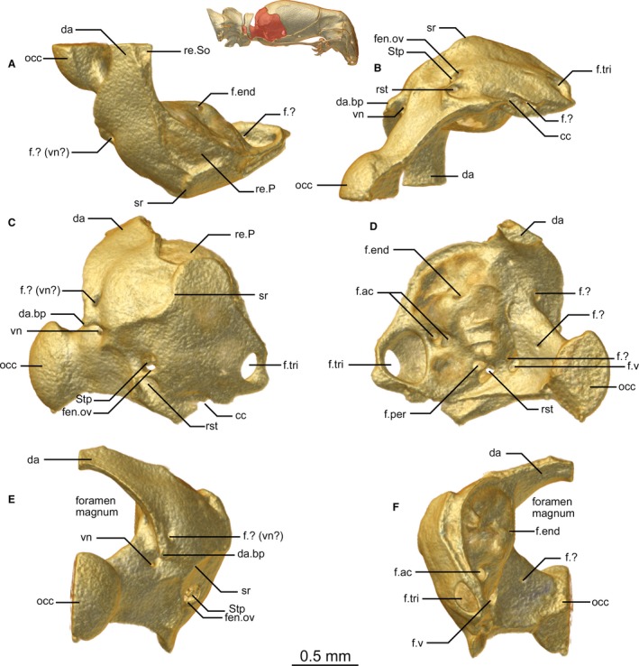
Exoccipital of Xenotyphlops grandidieri in (A) dorsal, (B) ventral, (C) lateral, (D) medial, (E) posterior, and (F) anterior view. cc, carotid channel; da, dorsal arch of the exoccipital; da.bp, bulbous process at base of the dorsal arch of the exoccipital; f.?, unknown foramen; f.ac, acoustic foramen; f.end, endolymphatic foramen; f.per, perilymphatic foramen; f.tri, trigeminal foramen; f.v, vagus foramen; fen.ov, fenestra ovalis; occ, occipital condyle; re.P, receiving surface for the parietal; re.So, receiving surface for the supraoccipital; rst, recessus scalae tympani; sr, sigmoid ridge; Stp, stapes; vn, vagus nerve.