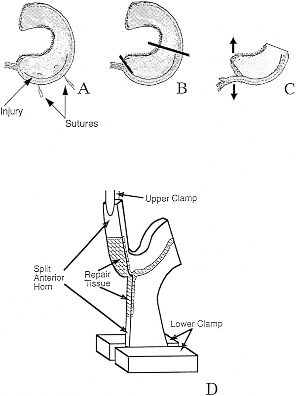
Figure 2.
Biomechanical testing of meniscus after peripheral injury and repair. After releasing the suture (A), the meniscus was split from the anterior horn to the beginning of the repaired tissue (B), thus producing two free ends (C). The posterior half of the meniscus was removed, and the two free ends of the anterior portion were inserted into clamps of a materials testing machine (D).

