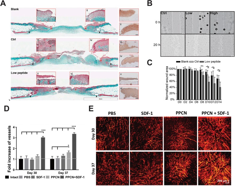
FIG. 2.
Advanced wound therapies aimed at accelerating wound closure by increasing the rate of re-epithelialization and angiogenesis. (a)–(d) Angiopoietin-1 derived peptide, QHREDGS, immobilized to a collagen-chitosan hydrogel. (a) Masson's trichrome stained cross sections of wounds in diabetic mice treated with no hydrogel, peptide free hydrogel, and QHREDGS conjugated hydrogel 14 days after wounding. Red arrowheads indicate the tips of the epithelial tongue, and black arrowheads indicate wounds edge. (Scale bar = 3 mm) Insets have been stained with pan-keratin to confirm epithelial tongue. (Scale bar = 50 μm). (b) Human epithelial keratinocytes seeded on no peptide control film, and films containing low (100 μM) and high (650 μM) concentration of QHREDGS peptides indicate that peptide containing films accelerate keratinocyte migration. (c) Quantification of the wound size reveals faster wound closure in wounds treated with the peptide hydrogel. Reproduced with permission from Xiao et al., Proc. Natl. Acad. Sci. U. S. A. 113(40), E5792 (2016). Copyright 2016 National Academy of Sciences.2 (d)–(e) SDF-1 entrapped in PPCN. (d) Quantification of blood vessel density following wound treatment with the PBS control, SDF-1 alone, PPCN alone, and SDF-1 entrapped in PPCN reveals a significant increase in vessel density. (e) Microangiography of the skin following treatment and healing on days 30 and 37. Reproduced with permission from Zhu et al., J. Controlled Release 238, 114 (2016). Copyright 2016 Elsevier.53

