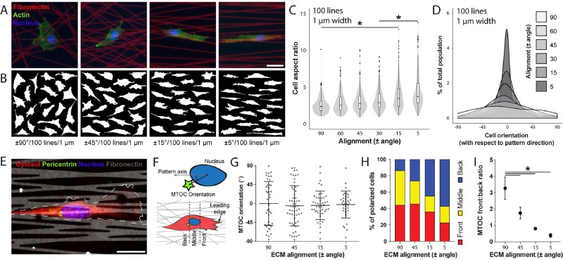
FIG. 3.
Increasing ECM alignment promotes an elongated uniaxial cell morphology and polarizes the cell. (a) Representative 3T3 fibroblasts seeded on Fn-patterns (red) of varying alignment with fixed line density and width (100 lines per pattern, 1 μm width), stained for F-actin (green) and nuclei (blue) with phalloidin-AlexaFluor488 and Hoechst33342, respectively (scale bar: 50 μm). (b) Cell outlines of 20 representative cells. (c) Aspect ratio and (d) cell orientation (angle between the long axis of the cell and the fiber alignment direction) of 3T3 fibroblasts as a function of ECM alignment, keeping line density constant at 100 lines per pattern and line width at 1 μm width (n ≥ 50 cells per condition, total of 515 cells analyzed). (e) Representative confocal image of a 3T3 fibroblast immunostained for pericentrin to localize the microtubule organizing center (MTOC) (red: cytosol, green: pericentrin, blue: nucleus, and grey: Fn555; scale bar: 25 μm). (f) Schematic indicating how MTOC orientation (angle between MTOC and the pattern axis using the centroid of the cell nucleus as a reference point) and position (front, middle, or back relative to the nucleus and leading edge of the cell) were determined. (g) MTOC orientation as a function of ECM alignment (n ≥ 35 cells per condition, total of 177 cells analyzed). (h) Percentage of all polarized cells with MTOC located in front, in the middle, or rearwards of the nucleus (n ≥ 60 cells per condition, total of 468 cells analyzed). (i) Ratio between cells with MTOC located in front of the nucleus to cells with MTOC located rearwards of the nucleus (* indicates a significant difference with p < 0.05).

