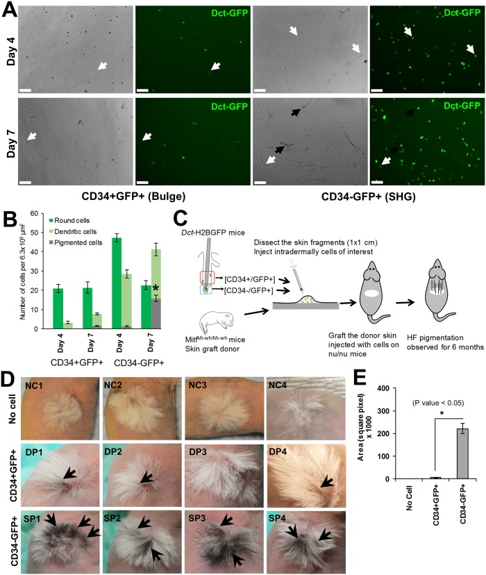
Fig 3. Distinct melanogenic properties of bulge and SHG melanocyte precursor cells of telogen HFs.
(A) In vitro melanocyte differentiation potential of CD34+GFP+ and CD34-GFP+ McSCs in melanocyte differentiation culture condition for day 4 (top two panels) or day 7 (bottom two panels). Dark arrow indicates pigmented cells and white arrow indicates dendritic but non-pigmented cells. Scale bars: 100 μm. (B) Quantification of bulge and SHG McSCs potential to produce pigmented melanocytes in melanocyte differentiation medium at day 4 and 7. *P ≤ 0.01. (C) Viable CD34+GFP+ and CD34-GFP+ McSCs isolated from Dct-H2BGFP mice were injected into skin fragments of neonatal MitfMi-wh/Mi-wh mice (P0 –P2). Injected skin fragments were engrafted onto nude mice to observe regeneration of HF pigmentation. (D) Skin grafts following reconstitution with CD34-GFP+ cells (SP) showed significantly greater regeneration of follicular pigmentation than grafts receiving CD34+GFP+ cells (DP). HF pigmented regions of the grafted skin are indicated by arrows. No cell (NC) injected grafted skin fragments were used as a negative control and showed no pigmented HFs. (E) Quantification of pigmented region of engrafted skin receiving CD34- McSCs, or CD34+ McSCs, or no cells. Area of pigmented region was calculated using Image J software. (* P Value < 0.05. by ANOVA; N = 5).

