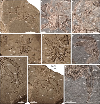
Fig. 1. Ventral and appendicular features in Callichimaera perplexa n. gen. n. sp., from the mid-Cretaceous of Colombia.

(A to C) Holotype IGM p881215. (A) Ventral view. (B) Close-up of sternal plates, sutures, and linea media (lm); the white arrow shows a spine in the third leg coxae. (C) Close-up of sternal crown, mandibles, and maxillipeds 2 and 3, the latter bearing a row of spines or crista dentata (cd) in the ischium. (D and E) Paratype IGM p881196. (D) Ventral view. (E) Close-up of sternum and sutures. (F) Paratype IGM p881185, showing details of the spanner-like claw. (G to I) Paratype IGM p881214. (G) Close-up of the large oar-like legs 2 and 3. (H) Ventral view showing the sternites, claws, legs, and pleon. (I) Close-up of the reduced and slender legs 4 and 5. Ba, basis; Ca, carpus; Da, dactylus; Pr, propodus; Ma, mandibula; Me, merus; Mxp2 and Mxp3, second and third maxillipeds, respectively; P1, claw or cheliped; P2 to P5, pereopods or legs 2 to 5; Po, pollex or fixed finger tip; Pl, pleon or “abdomen”; S1 to S7, sternites 1 to 7; 4/5–6/7, sternal sutures. All specimens were photographed dry; specimens in (A), (D), (E), (G), and (H) were coated with ammonium chloride; specimens in (B), (C), (F), and (I) were uncoated. Photos by J.L.
