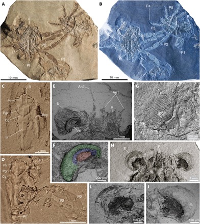
Fig. 2. Dorsal, frontal, and ocular features in Callichimaera perplexa n. gen. n. sp., from the mid-Cretaceous of Colombia.

(A and B) Paratype MUN-STRI 27044-02, showing three specimens in dorsal view (left), ventral view (right), and an isolated dorsal carapace (bottom center). (A) Color image. (B) Inverted color image highlighting details of the carapace outlines, claws and legs, and large eyes. (C) Paratype IMG p881203, dorsal view, showing details of the carapace grooves and ridges and the lack of true orbits. (D) Paratype IMG p881218, dorsal view, showing details of dorsal carapace, claws, and large oar-like legs 2 and 3. (E and F) Paratype IGM p881209a. (E) Scanning electron microscope (SEM) image showing details of large compound eye, optical lobe, rostrum (R), and two pairs of short antennae between the eyes. (F) SEM showing a close-up of the optical lobe. (G) Paratype IGM p881220, SEM of large eye preserving mostly hexagonal facets in hexagonal arrangement (left box), although the proximal cornea bears squarish facets in orthogonal packing (right box). (H to J) Paratype IGM p881208, ventral view, (H) showing the large eyes and bifid rostrum, (I) SEM of ventral right eye, and (J) SEM of ventral left eye. An1, antenna 1 or antennula; An2, antenna 2; Bcg, branchio-cardiac groove; Cg, cervical groove; E, compound eye; Lr, longitudinal ridge; Ol, optical lobe; Pfr, postfrontal longitudinal ridge; Pgr, protogastric longitudinal ridge. All specimens were photographed dry; specimens in (C) and (D) were coated with ammonium chloride; specimens in (A), (B), and (E) to (J) were uncoated. Photos by J.L.
