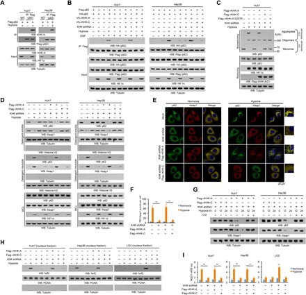
Fig. 1. KHK-A is required for oxidative stress–enhanced p62 oligomerization and Nrf2 activation in HCC cells.

(A to D and G and H) Immunoblot analyses were performed with the indicated antibodies. (A) Huh7 and Hep3B cells expressing Flag-p62 were treated with or without hypoxia for 6 hours in the presence of 10 μM chloroquine (CQ; a lysosome inhibitor). Immunoprecipitation analyses with an anti-Flag antibody were performed. Hif 1α, hypoxia-inducible factor 1α; WB, Western blot; IP, immunoprecipitation; IgG, immunoglobulin G. (B) Huh7 and Hep3B cells with or without expressing KHK short hairpin RNA (shRNA) and with or without reconstituted expression of the indicated proteins were transfected with vectors expressing Flag-p62 and HA-p62 and treated with or without hypoxia for 6 hours in the presence of the lysosome inhibitor CQ (10 μM). After incubation with the reversible cross-linking agent DSP (0.4 mg/ml) for 2 hours, the cells were lysed in a buffer containing 1% SDS to solubilize all proteins. The lysates were subjected to immunoprecipitation analyses with an anti-Flag antibody after diluting SDS to 0.1%. (C) Huh7 cells with or without expressing KHK shRNA and with or without reconstituted expression of the indicated proteins were treated with or without hypoxia for 6 hours and lysed and analyzed by reducing (containing 2.5% β-mercaptoethanol) and nonreducing SDS–polyacrylamide gel electrophoresis (SDS-PAGE) to detect p62 aggregation. (D) Huh7 and Hep3B cells with or without expression of KHK shRNA were reconstituted with or without expression of the indicated KHK proteins. After stimulation with or without hypoxia for 6 hours in the presence of the lysosome inhibitor CQ (10 μM), the cells were lysed in a lysis buffer with 1% Triton X-100. The insoluble fraction was lysed in a lysis buffer with 1% SDS. WCL, whole-cell lysate. (E and F) Huh7 cells with or without KHK shRNA expression and with or without reconstituted expression of Flag-tagged rKHK-A or rKHK-C were stimulated with or without hypoxia for 6 hours. Immunofluorescent analyses were performed with the indicated antibodies (E). The numbers of puncta in 100 cells were counted and quantified. Data are shown as means ± SD of 100 cells per group. A two-tailed Student’s t test was used. **P < 0.01 (F). (G) Huh7 and Hep3B cells expressing KHK shRNA with or without reconstituted expression of the indicated proteins were treated with or without hypoxia and lysosome inhibitor CQ (10 μM) for the indicated periods of time. (H) The indicated cells with or without expressing KHK shRNA and with or without reconstituted expression of the indicated proteins were treated with or without hypoxia for 12 hours. The nuclear fractions were prepared. PCNA, proliferating cell nuclear antigen. (I) The indicated cells with or without expressing KHK shRNA and with or without reconstituted expression of the indicated proteins were transfected with quinone oxidoreductase 1 (NQO1)–ARE-luc and pRL-TK (Renilla luciferase control reporter vector) plasmids. Starting at 18 hours after transfection, cells were treated with or without hypoxia for 12 hours and harvested for luciferase activity analyses. The data are presented as means ± SD from triplicate samples. **P < 0.01.
