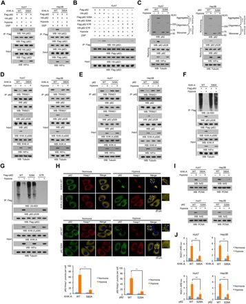
Fig. 4. KHK-A–mediated p62 S28 phosphorylation is required for oxidative stress–enhanced p62 oligomerization and Nrf2 activation.

(A to G and I) Immunoprecipitation and immunoblot analyses were performed with the indicated antibodies. (A) Huh7 and Hep3B cells with or without knock-in of KHK-A S80A expression were transfected with the vectors expressing Flag-p62 and HA-p62 and treated with or without hypoxia for 6 hours in the presence of the lysosome inhibitor CQ (10 μM). After incubation with DSP (0.4 mg/ml) for 2 hours, the cells were lysed in a buffer containing 1% SDS to solubilize all proteins. The lysates were subjected to immunoprecipitation analyses with an anti-Flag antibody after diluting SDS to 0.1%. (B) Huh7 cells expressing the indicated p62 proteins were treated with or without hypoxia for 6 hours in the presence of the lysosome inhibitor CQ (10 μM). After incubation with DSP (0.4 mg/ml) for 2 hours, the cells were lysed in a buffer containing 1% SDS to solubilize all proteins. The lysates were subjected to immunoprecipitation analyses with an anti-Flag antibody after diluting SDS to 0.1%. (C) Huh7 and Hep3B cells with or without knock-in expression of p62 S28A were treated with or without hypoxia for 6 hours. The whole-cell lysates were analyzed by reducing and nonreducing SDS-PAGE to detect p62 aggregation. (D and E) Huh7 and Hep3B cells with or without knock-in expression of KHK-A S80A (D) or p62 S28A (E) were stimulated with or without hypoxia for 6 hours in the presence of the lysosome inhibitor CQ (10 μM). (F) Huh7 cells with or without knock-in of KHK-A S80A expression were treated with or without hypoxia for 6 hours in the presence of the lysosome inhibitor CQ (10 μM). (G) Endogenous p62-depleted Huh7 cells with or without reconstituted expression of WT Flag-rp62, rp62 S28A, or rp62 K7R were treated with or without hypoxia for 6 hours in the presence of the lysosome inhibitor CQ (10 μM). (H) Huh7 cells with or without knock-in of KHK-A S80A (top) or p62 S28A (bottom) expression were stimulated with or without hypoxia for 6 hours. Immunofluorescence analyses were performed with the indicated antibodies. Numbers of puncta in 100 cells were counted and quantified. Data are shown as means ± SD of 100 cells per group. A two-tailed Student’s t test was used. **P < 0.001. (I) Huh7 and Hep3B cells with or without knock-in of KHK-A S80A (top) or p62 S28A (bottom) expression were treated with or without hypoxia for 12 hours. The nuclear fractions were prepared. (J) Huh7 and Hep3B cells with or without knock-in of KHK-A S80A (top) or p62 S28A (bottom) expression were transfected with NQO1-ARE-luc and pRL-TK plasmids. After treatment with or without hypoxia for 12 hours, the cells were harvested for luciferase activity analyses. The data are presented as means ± SD from triplicate samples. **P < 0.001.
