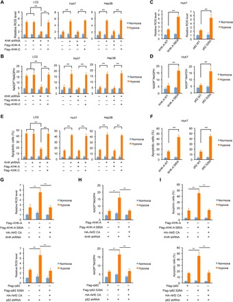
Fig. 5. p62 S28 phosphorylation reduces ROS production and promotes cancer cell survival.

The data are presented as means ± SD from three independent experiments. **P < 0.001. A two-tailed Student’s t test was used. (A, B and E) LO2, Huh7, and Hep3B cells with or without expression of KHK shRNA were reconstituted with or without expression of the indicated KHK proteins. After treatment of the cells with or without hypoxia for 36 hours, ROS levels (A), intracellular NADP+ and NADPH levels (B), and the percentages of apoptotic cells (E) were measured. (C, D, and F) Huh7 cells with or without knock-in of KHK-A S80A (left) or p62 S28A (right) expression were treated with or without hypoxia for 36 hours. ROS levels (C), intracellular NADP+ and NADPH levels (D), and the percentages of apoptotic cells (F) were measured. (G to I) Huh7 cells with reconstituted expression of the indicated KHK-A proteins (top) or p62 proteins (bottom) were stably transfected with or without HA-tagged constitutively active Nrf2 and treated with or without hypoxia for 36 hours. ROS levels (G), intracellular NADP+ and NADPH levels (H), and the percentages of apoptotic cells (I) were measured.
