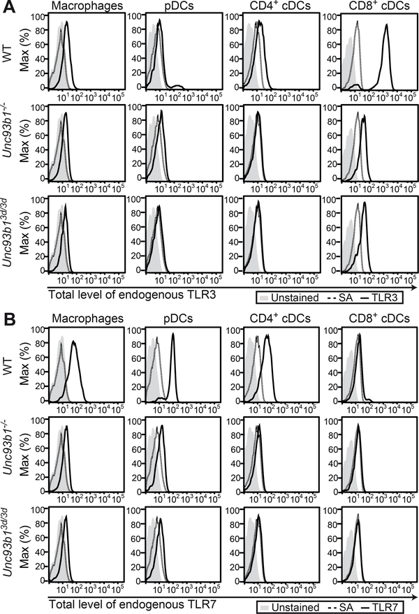
Figure 7. Protein expression of endogenous TLRs is severely reduced in Unc93b1−/− and Unc93b13d/3d mice.
(A and B) Splenocytes from WT, Unc93b1−/− and Unc93b13d/3d mice were isolated and stained for different immune cell subsets, namely macrophages, pDCs, CD4+ and CD8+ cDCs. Upon permeabilization, cells were stained with (A) biotinyated anti-TLR3 antibody (clone PAT3) (Murakami et al., 2014) or (B) biotinylated anti-TLR7 antibody (clone A94B10) (Kanno et al., 2013) coupled to fluorescently labeled streptavidin (SA) and analyzed by flow cytometry. Dotted histograms represent negative controls stained with fluorescently labeled SA alone. Data are representative of two independent experiments. Please see Figure S4 for gating strategies.

