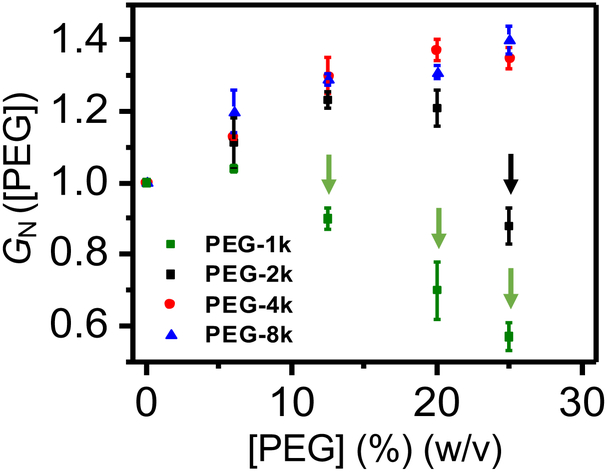
Figure 2: Normalized conductance of the αHL protein pore, GN ([PEG]).
Here, GN ([PEG]) = (G([PEG]))/(G([0])), where G([0]) was the measured unitary conductance in 300 mM KCl, 10 mM Tris⋅HCl, pH 7.4, and G([PEG]) was the measured unitary conductance in the presence of PEG-1k (green squares), PEG-2k (black squares), PEG-4k (red circles) or PEG-8k (blue triangles) added symmetrically to both sides of the chamber. The vertical arrows highlight cases in which the normalized conductance of the αHL protein pore in the presence of PEG, GN, is smaller than 1. For these cases, PEGs are easily-penetrating polymers. The applied transmembrane potential was +80 mV.

