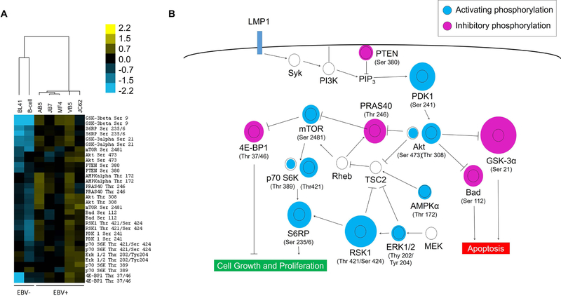
Figure 1: EBV-positive B cell lines from PTLD patients have constitutive over-activation of the PI3K/Akt/mTOR pathway.
(A) Cell lysates were prepared from B cells isolated from a healthy donor (first column), an EBV-negative Burkitt’s lymphoma line (BL41, second column), and five EBV-positive B cell lymphoma samples taken from patients with PTLD (AB5, JB7, JC62, MF4, VB5). The relative phosphorylation levels of the measured nodes for each sample is represented as a heat map, using hierarchical clustering based on Euclidean distances. The two EBV-negative cell samples are clustered separately from the EBV-positive lymphoma cell lines based on differences in the phosphorylation pattern. (B) The relative phosphorylation levels of each node is depicted within the context of the PI3K signaling pathway. The relative diameter of the blue or purple circle to the black ring is proportional to the fold-difference in average phosphorylation levels of the five EBV+ lymphoma cell lines compared to the normal B cell control. The constitutive phosphorylation patterns of the EBV+ lymphoma cell lines overall promote cell proliferation and inhibit apoptotic checkpoints.

