Fig. 3.

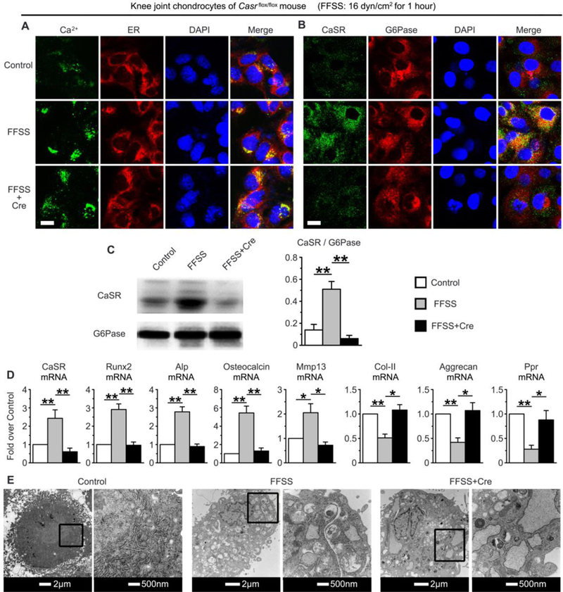
The impact of Casr gene knockout on FFSS-induced changes in (A) Ca2+ loading, (B, C) CaSR localization in ERs, (D) expression of cell differentiation markers, and (E) organelle swelling in primary chondrocytes cultured from knee joints of Casrflox/flox mice. Induction of gene KO was performed by transduction of Cre protein in chondrocyte cultures for 24 hrs before the application of FFSS. (A) Dual-fluorescence staining of Ca2+ and ERs by Fluo-8 (in green) and ER-tracker (in red) respectively, showed inability of Casr KO to prevent the FFSS-induced Ca2+ loading in ERs. (B) Dual-fluorescence staining and (C) immunoblotting of CaSR (in green) and G6Pase (in red) showed the ability of Casr KO to abrogate the FFSS-induced CaSR localization in ERs. Bar=5μm. (D) mRNA expression profiles by qPCR showed the ability of Casr KO to block the impact of FFSS on the expression of early and terminal differentiation markers. Values are presented as the mean±SD. **p<0.01, *p<0.05 between groups as specified by top horizontal bars in each panel. (E) TEM images showed the inability of Casr KO to affect the FFSS-induced swelling of mitochondria and ERs. n=4 separate cultures.
