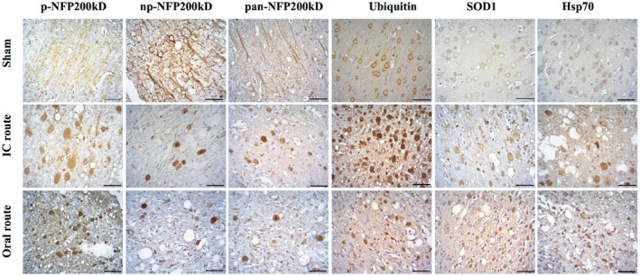
Figure 2.

Images showing rat axonal swellings (spheroids) surrounding T. solium cysticercus. Spheroids were detected using IHC to neurofilament (phosphorylated (p‐NFp), non‐phosphorylated (np‐NFp) or pan), ubiquitin, SOD1 or Hsp70 antibodies. No spheroids were found in sham animals. The images show spheroids located in cortical areas adjacent to the cysticercus for the intracranial (IC) and oral routes of infection but were also found in the hippocampus (data not shown). Spheroids were more clearly observed using NFp than with np‐NFp or pan‐NFp antibody. Ubiquitin and SOD1 in sham sections were restricted to the cytoplasm as expected. In both IC and oral routes of infection, ubiquitin and SOD1 spheroids were scattered in the cortex and associated with spongy changes. Hsp70 spheroids were fewer compared to other markers. Scale bars = 50 μm.
