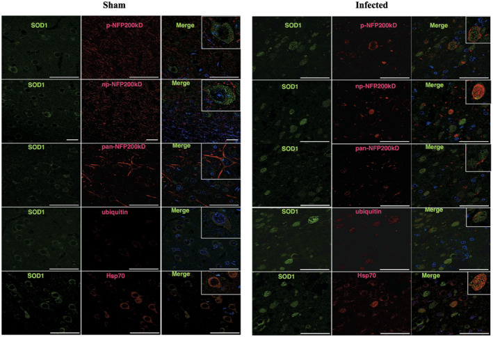
Figure 4.

Spheroids shown by confocal imaging. Sham sections showed the normal distribution of SOD1, neurofilament forms, ubiquitin and Hsp70, with no spheroids seen. Infected brains (intracranial route) show most neurofilament protein (p‐NFP200, np‐NFP200KDa and pan‐NFP200KDa) forms do not overlap with SOD1. However, as is shown in SOD1 and p‐NFP200KDa immunofluorescence, SOD1 spheroids can be surrounded by or can coexist with NFp. SOD1 and ubiquitin double labeling shows that spheroids share a similar distribution as well as SOD1 and Hsp70 spheroids.
