
Figure 2. The majority of foam cells in ApoE−/− mice fed a Western diet are non-leukocyte in origin.
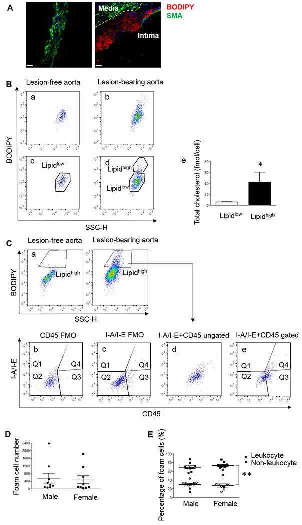
(A) Representative immunostaining of lesion-free descending aorta (left panel) and lesion-bearing aortic arch (right panel) of 8-week-old ApoE−/− mice after 6 weeks of WD feeding. Foam cells were labeled with BODIPY (red). The yellow dashed line indicates the internal elastic lamina separating the medial layer exhibiting absence of lipid staining and high levels of SMA (green), and intimal layer containing foam cells (red) and much lower levels of SMA. (B) Histogram identifying foam cells from the aortic arch. Nucleated single events from lesion-bearing aortas (b) have a cell population with higher BODIPY intensity comparing to lesion-free descending aortas (a). Cells from lesion-free aortas were gated as the Lipidlow population (c), and cells above this threshold in atherosclerotic aortic arch were gated as the Lipidhigh population (d). Cholesterol content in cells in the Lipidhigh population was much higher than cells in the Lipidlow population. N=4 using cells sorted from 4 different animals, *P < 0.05 using paired one-sided Student’s t test (e). (C) Lipidhigh cells in the aortic arch of another mouse were identified by comparison to lipid content in lesion-free descending aorta of the same mouse (a), and further separated by their expression of CD45 and I-A/I-E (e). A small aliquot of the same cell suspension was stained in the absence of either CD45 (“fluorescence minus one”, FMO, b) or I-A/I-E (c) antibodies as negative controls to set the gates positive for CD45 (Q3 and Q4) and I-A/I-E (Q1 and Q4). Lipidhigh foam cells were separated by I-A/I-E and CD45 expression at the same time based on the position of the antibody-negative control gates (d and e). (D) Foam cell numbers in the lesion-bearing aortas of individual male and female mice. N=9 mice/group. No statistical differences were found by Mann-Whitney U test between the sexes. (E) Percentage of non-leukocyte foam cells negative for both macrophage markers (Q2) and leukocyte-derived foam cells (Q3+Q4) out of total BODIPY+ events in male and female ApoE−/− mice. N=9 mice/group, **P < 0.01 using 2-way ANOVA with Bonferroni post hoc comparisons.