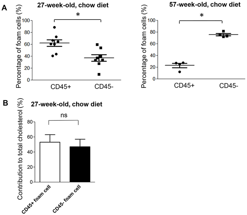
Figure 4. Percentage of non-leukocyte- and leukocyte-derived foam cells in spontaneous atherosclerosis of ApoE−/− mice and their contribution to cholesterol accumulation in arteries.

(A) CD45− and CD45+ foam cells in the aortic arch were quantified in male 27- (left panel, n=8) and 57-week-old (right panel, n=4) ApoE−/− mice fed a chow diet. *P < 0.05 using a Student’s t test (left panel) or Mann–Whitney U test (right panel). (B) CD45+ and CD45− foam cells were separated by flow cytometry from 27-week-old mice. Sorted foam cells from 1-3 animals were pooled to obtain sufficient cells for mass determination of cholesterol content (n=3 data sets from a total of 8 animals). There was no significant differences in the contribution of CD45+ and CD45− foam cells to total cholesterol content in isolated foam cells by Wilcoxon matched pairs sign rank test.
