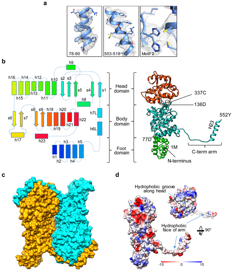
Figure 2: Structure of the NS1 monomer.
(a) Superposition of the cryoEM densities (gray mesh) and their corresponding atomic models (ribbon and sticks) for three selected regions of NS1 illustrating the quality of the cryoEM densities that supports atomic modeling based on amino acid side chains. (b) Secondary structure schematic and domain architecture (left) mapped to the atomic model (shown as ribbon diagram to the right) of an NS1 Monomer. (c) Space-filling surface rendering of an NS1 dimeric building subunit of both tubular forms. (d) Coulombic surface rendering of the NS1 monomer showing the C-terminal arm handshake. Note the hydrophobic groove in the head domain and the hydrophobic inner surface of the C-terminal arm.

