Fig. 3:
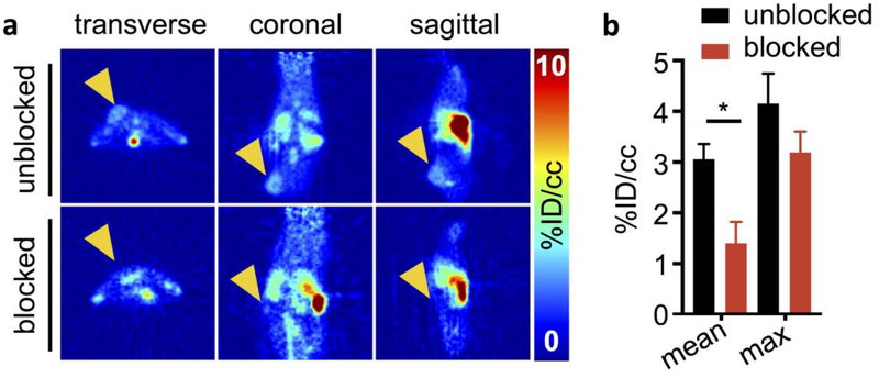
In vivo PET imaging of [18F]FE-OTS964 in U87 tumor-bearing NSG mice. a PET images (left to right: transverse, coronal, and sagittal) centered on the tumor in top (unblocked) mice injected with [18F]FE-OTS964 and bottom (blocked) mice injected with [18F]FE-OTS964 + 5 mg/kg OTS514. Arrows indicate tumor location. b VOI quantification of PET scans. Statistically significant differences were seen in mean voxel %ID/cc between blocked (black circles) and unblocked (red squares) mice. *Significant at p<0.05.

