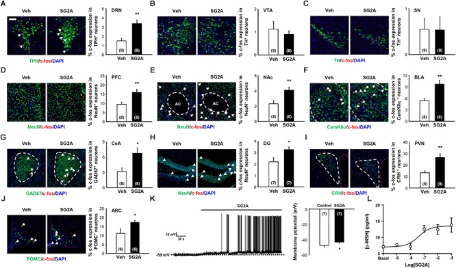
Figure 4.

Neurons responding to SG2A. Immunohistochemistry for c-fos+ (red) neurons in mice administered SG2A or vehicle (Veh) for 1–2 h. The Percentage of cells double immunopositive for c-fos and TPH (serotonergic neurons) in the DRN (A), TH (dopaminergic neurons) in the VTA (B) and SN (C), NeuN in PFC (D) and NAc (E), CamKIIα in BLA (F), GAD67 in CeA (G), NeuN in the DG of the hippocampus (H), CRH in PVN (I), and POMC in ARC (J) were counted (3–4 slices were counted per mouse). (K) Depolarization of POMC neuron membrane potential in response to applications of SG2A (left); summary of acute effects of SG2A on the membrane potentials of responsive POMC neurons (right). (L) α-MSH secretion in cultured POMC neurons in response to SG2A. Data are presented as means ± SEMs; ∗P < 0.05 and ∗∗P < 0.01 vs. Veh. Numbers in parentheses indicate the numbers of animals used for each group. Scale bar, 50 μm.
