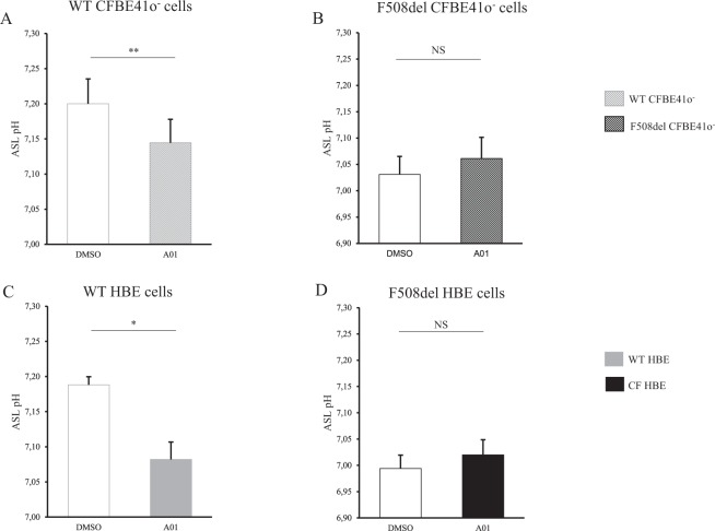
Figure 4.
Airway surface liquid pH measurements upon SLC26A4 inhibition in WT and F508del bronchial epithelia. CFBE41o− cells and Human Bronchial Epithelial (HBE) primary cells were incubated with DMSO vehicle (empty bars) or specific pendrin inhibitor A01 (filled bars) for 6 h. ASL pH was measured after apical addition of 50 µL Ringer’s solution (25 mM HCO3−, 5% CO2). DMSO induces a slight ASL acidification in both WT and F508del cells reaching a decrease of 0.2 pH units after 6 h incubation. (A) WT CFBE41o− cells. pH = 7.20 ± 0.04 (DMSO) vs. pH = 7.14 ± 0.03 (A01), p = 0.008. (B) F508del CFBE41o− cells. pH = 7.03 ± 0.03 (DMSO) vs. pH = 7.06 ± 0.04 (A01), NS. (C) WT HBE primary cells. pH = 7.19 ± 0.01 (DMSO) vs. pH = 7.08 ± 0.02 (A01), p = 0.04. (D) F508del HBE primary cells. pH = 6.99 ± 0.03 (DMSO) vs. pH = 7.02 ± 0.03 (A01), NS. For all conditions, n = 3 in triplicate. Data are presented as mean ± SEM. Statistical significance from unpaired nonparametric Wilcoxon test. *p < 0.05; **p < 0.01; NS: non significant.

