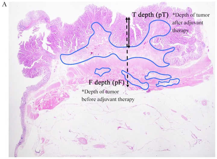
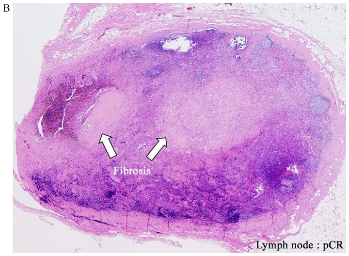
Figure 1.
Hematoxylin and eosin staining. (A) Low magnification view of a hematoxylin and eosin-stained section (magnification, ×2). The F depth (fibrosis) and T depth are the depths of the tumor from the mucosal surface prior to and following adjuvant therapy, respectively. Areas of fibrosis were surrounded with a blue outline. (B) Example of pCR for a lymph node. pCR was defined as the absence of histological evidence of vital tumor cells at the site of the primary tumor or lymph nodes and the existence of fibrosis (magnification, ×2). pCR, pathological complete response.


