Fig. 10.
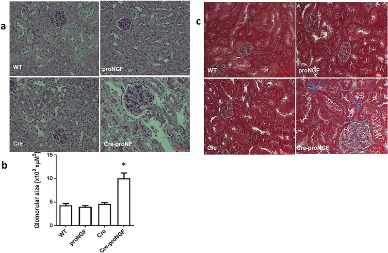
EC-proNGF overexpression induces glomerular enlargement and fibrosis. Histopathological changes Including renal fibrosis and glomerular sclerosis was assessed by Masson’s trichrome staining and PASH staining after 8-weeks of EC-proNGF overexpression. (a & b) Representative and statistical analysis using One-Way ANOVA of PASH-stained kidney sections from Cre-proNGF mice showed glomerular expansion and mesangial expansion compared to control groups (*p < 0.001 versus control, n = 4). (c) Masson Trichrome staining showed increased glomeruli enlargement and the interstitial fibrosis (blue) in kidney sections from Cre-proNGF mice compared to other controls (n = 4). Scale bar = 100 μM.

